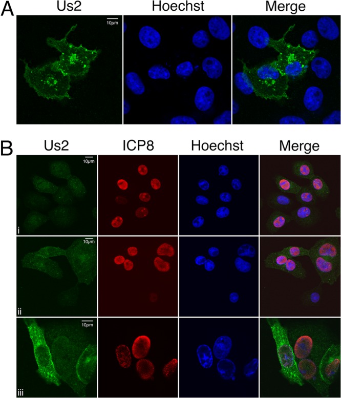
Fig 2.

Subcellular localization of HSV-2 Us2. (A) HSV-2 Us2 localizes to membranes in transfected Vero cells. Shown is a representative image of Vero cells transfected with a plasmid encoding HSV-2 Us2 and stained with a polyclonal antiserum specific for HSV-2 Us2 (green) at 24 h posttransfection. Nuclei were detected using the DNA stain Hoechst 33342. (B) Kinetics of HSV-2 Us2 expression and localization in infected Vero cells. Shown are representative images of Vero cells infected with HSV-2. Cells were fixed at 6 h (i), 10 h (ii), or 18 h (iii) after infection and were stained for Us2 (green) or the HSV-2 nuclear protein ICP8 (red). Nuclei were stained with Hoechst 33342 (blue). Images of stained cells were captured by confocal microscopy.
