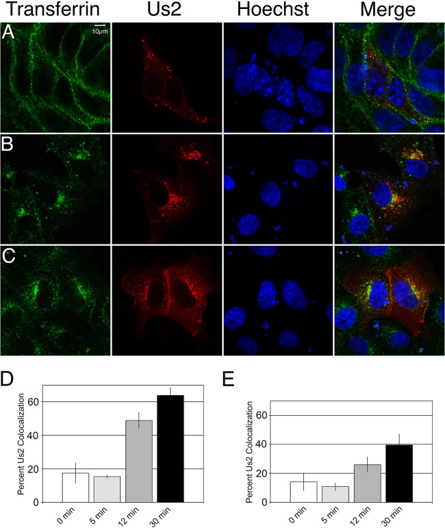
Fig 7.
A subset of Us2 colocalizes with endosomal vesicles. (A to C) Vero cells transfected with an mCherry–HSV-2 Us2 expression plasmid were incubated with Alexa Fluor 647-conjugated transferrin for 30 min at 4°C, washed, and shifted to 37°C for 5 min (A), 12 min (B), or 30 min (C) prior to fixation. The transferrin signal is shown in green. The Us2 signal is red. Nuclei were detected using the DNA stain Hoechst 33342 (blue). Representative images are shown. (D) Quantification of transferrin colocalization with Us2 puncta in cells transfected with mCherry–HSV-2 Us2. The data are presented as percentages of Us2-containing puncta that are also positive for transferrin. A total of 12 fields of cells and 298 Us2 puncta were analyzed. Error bars represent the standard errors of the means observed for differences between different fields of cells. (E) Quantification of transferrin colocalization with Us2 puncta in HSV-2-infected cells at each time point. The data are presented as percentages of Us2-containing puncta that are also positive for transferrin. Us2 puncta were identified by indirect immunofluorescence confocal microscopy using a polyclonal antiserum against Us2 and an Alexa Fluor 588-conjugated secondary antibody. A total of 30 fields of cells and 938 Us2 puncta were analyzed. Error bars represent the standard errors of the means observed for differences between different fields of cells at each time point.

