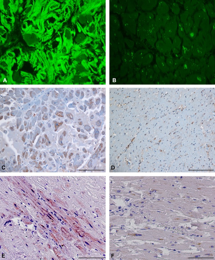
Fig 3.
Amyloid and protease-resistant PrP are present in FFPE amyloid heart tissue. (A) Section of heart tissue from sample AHD1FFPE stained with ThioS. Significant ThioS immunofluorescence indicative of amyloid can be seen surrounding the cells. (B) Section of heart tissue from sample NHt2FFPE stained with ThioS. No significant immunofluorescence is present. (C) Section of heart tissue from sample AHD1FFPE stained with the anti-PrP antibody PV30. Antibody reactivity is represented by the brown stain over the cells, indicating the presence of PrP. The pale blue stain surrounding the cells is amyloid. (D) Section of heart tissue from sample NHt2FFPE stained with the anti-PrP antibody PV30. Antibody reactivity is localized primarily to blood vessels and blood cells. (E) Section of heart tissue from sample AHD1FFPE stained with the anti-PrP antibody PV30 after treatment with PK. Antibody reactivity is represented by the brown stain over the cells and indicates the presence of protease-resistant PrP. (F) Section of heart tissue from a patient diagnosed with amyloid heart disease (AHD1FFPE). The section was stained with PV30 preabsorbed with a peptide containing the PV30 antibody epitope. No positive signal was seen. For all panels, the scale bar represents 100 μm.

