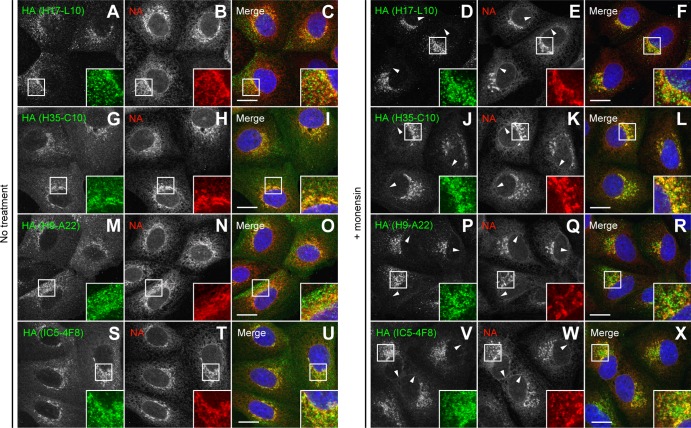
Fig 3.
Reactivity of various anti-HA MAbs assayed by immunofluorescence microscopy. MDCK cells were infected with IAV PR8 in the absence (no treatment) or presence of 10 μM monensin as described in the legend to Fig. 2E to V. HA was labeled on fixed and permeabilized cells with specific MAbs to the HA Ca (H17-L10, A to F), Cb (H35-C10, G to L), Sa (H9-A22, M to R), and Sb (IC5-4F8, S to X) antigenic sites (green channel). NA was detected with rabbit polyclonal Abs (red channel). DNA was labeled with DAPI (blue channel). Stained cells were examined by confocal fluorescence microscopy. Bars, 10 μm. Arrowheads point to NA colocalization with HA monomers on the nuclear envelope (ER) of cells labeled with IC5-4F8.

