Fig 2.
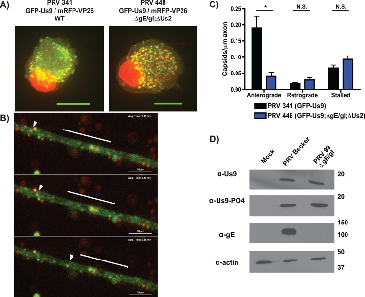
gE/gI are required for efficient axonal sorting and anterograde transport of viral particles. (A) Dissociated SCG cultures were infected with PRV 341 (expresses GFP-Us9 and the capsid protein mRFP-VP26) or PRV 448 (expresses GFP-Us9 and mRFP-VP26 and is null for gE, gI, and Us2). Infected cells were fixed at 8 hpi and visualized by confocal microscopy. Each image represents a maximum-intensity projection. (B) Live-cell imaging of anterograde transport of virions at 8 hpi with PRV 448, showing GFP-Us9 and mRFP-VP26 channels over time. The arrowhead indicates the position of a dually labeled viral particle at different time points. In each frame, the arrow above the axon indicates the direction of anterograde movement. (C) Quantification of anterograde transport of capsids in axons infected with PRV 341 or PRV 448. Ten axons from two biological replicates were imaged for 3 min each, and the numbers of anterograde, retrograde, and stalled capsids were manually quantified. The number of viral particles counted in each axon was normalized to axon length. Error bars indicate standard errors of the means (SEM). *, P < 0.001; N.S., not significant. (D) Differentiated PC12 cells were infected with PRV Becker or PRV 99 (null for gE and gI), lysed at 12 hpi, and subjected to Western blot analysis using the indicated antibodies.

