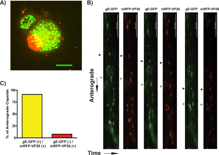
Fig 5.
Visualization of gE-GFP in PRV-infected SCG neurons. (A) Confocal microscopy of an SCG cell body infected with PRV 199 (expresses gE-GFP and mRFP-VP26) at 8 hpi. (B) Live-cell epifluorescence microscopy imaging of SCG axons was performed at 8 hpi for 3 min per axon. GFP and mRFP channels are shown sequentially. Two anterograde-directed puncta, each representing enveloped viral particles, are highlighted with black and green triangles, respectively. (C) Anterograde-directed mRFP-VP26-positive punctate structures were scored for colocalization with gE-GFP. A total of 65 puncta were counted across 5 movies, representative of 2 biological replicates.

