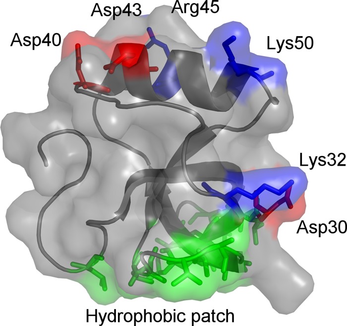
Fig 1.

Three-dimensional structure of Trichoderma reesei HHBI (Protein Data Bank accession number 2FZ6). Basic and acidic residues are annotated and colored blue and red, respectively. The protein binds to hydrophobic substrates through the hydrophobic patch (shown in green).
