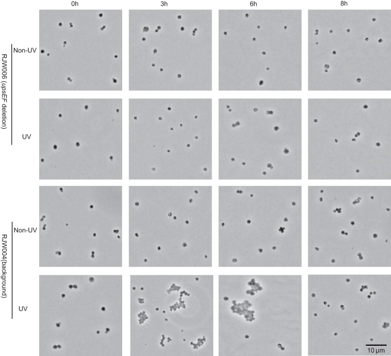
Fig 6.
Microscopy analysis of RJW006 (upsEF deletion mutant) and RJW004 (background strain) after UV treatment or without UV treatment. Ten microliters of S. islandicus cells was fixed on a Gelrite-coated microscope slide and then analyzed using Olympus BX60 (phase contrast). Representative micrographs of cells at 0 h, 3 h, 6 h, and 8 h of incubation after UV treatment or without UV treatment are shown.

