Fig 1.
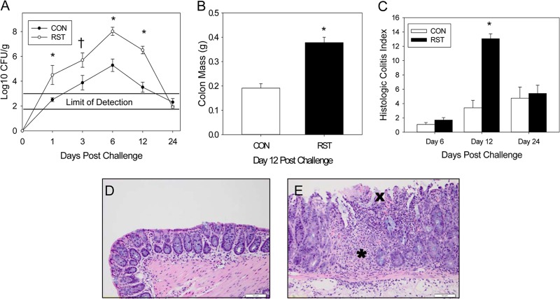
Stressor exposure enhances C. rodentium-induced infectious colitis. Mice were restrained for 1 day prior to oral challenge with 3 × 10⁸ CFU of C. rodentium. Restraint continued for 6 days postchallenge. (A) To determine the extent of colonization by C. rodentium, stool samples were collected on days 0, 1, 3, 6, 12, and 24 postchallenge. The pathogen was enumerated via the pour plate method. Exposure to restraint stress significantly increased C. rodentium colonization compared to that in CON mice. *, P < 0.005 for RST versus CON groups on indicated days postchallenge, as assessed with Student's t test with the Bonferroni correction factor as a post hoc test; †, P = 0.07 for RST versus CON groups on day 3 postchallenge, as assessed with Student's t test. Sample sizes were as follows: day 1, n = 11 (CON) and n = 5 (RST); days 3, 6, and 24, n = 12 (CON) and n = 5 (RST); and day 12, n = 16 (CON) and n = 8 (RST). (B) Colons were removed on day 12 and weighed without contents. Exposure to prolonged restraint significantly increased the weight of the colonic tissue compared to that of CON mice. *, P < 0.0001 for CON versus RST groups as assessed using ANOVA. Sample sizes were as follows: n = 10 (CON) and n = 5 (RST). (C) On days 6, 12, and 24 postchallenge, colons were removed, fixed in formalin, and subsequently embedded in paraffin. Colons were sectioned and stained with hematoxylin and eosin in order to visualize and score the pathology present in each sample. Stressor exposure significantly increased the total pathology scores. *, P < 0.00001 between RST and CON groups on day 12 postchallenge, as assessed using Student's t test with the Bonferroni correction as a post hoc test. Sample sizes were as follows: day 6, n = 6 (CON) and n = 3 (RST); day 12, n = 16 (CON) and n = 8 (RST); and day 24, n = 12 (CON) and n = 6 (RST). (D) Representative image of H&E-stained colon from a CON mouse on day 12 postchallenge. (E) Representative image of H&E-stained colon from an RST mouse on day 12 postchallenge. The asterisk marks a region with significant inflammatory cell infiltration, whereas the “X” marks regional ulcerations in the surface epithelium.

