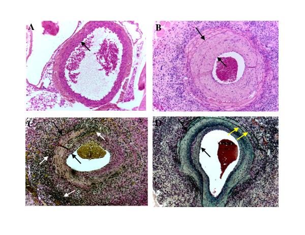
Figure 2.
Neointima formation in the aortas of MCMV infected IFN-γR-/- mice 2 months after whole body irradiation. A. H&E staining of abdominal aorta collected from uninfected IFN-γR-/- mouse 2 months after whole body irradiation. Arrow points to the intima. B. H&E staining of a large abdominal vessel collected from MCMV infected IFN-γR-/- mouse 4 months after infection and 2 months after whole body irradiation. Space between arrows indicates the thickness of the neointima C. Verhoeff-van Gieson stain of a large abdominal vessel collected from MCMV infected IFN-γR-/- mouse 4 months after infection and 2 months after whole body irradiation. Verhoeff-van Gieson stain: muscle (brown-yellow), collagen (red), cell cytoplasm (yellow), nuclei (blue-black). Focal transmural necrotizing vasculitis is seen. . Space between black arrows indicate intimal thickening and white arrows indicate focal adventitial inflammation and fibrosis. D. Movat staining of a large abdominal vessel collected from MCMV infected IFN-γ R-/- mouse 4 months after infection and 2 months after whole body irradiation. Movat stain: elastica (black), collagen and reticular fibers (yellow), mucosubstance (blue-green), red blood cells (red). Space between black arrows indicates the thickness of the neointima. Yellow arrows indicate normal external and internal elastica. Objective magnification x 20.

