
Fig 3.
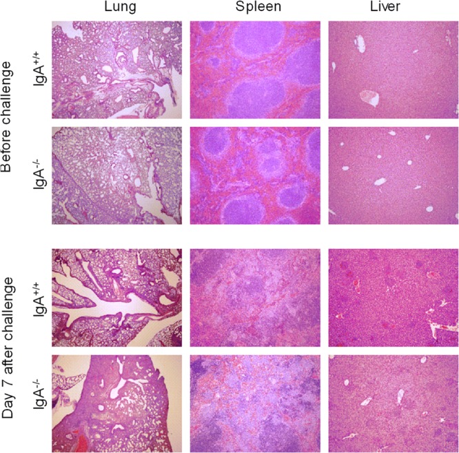
Histology of organs from naive and F. tularensis LVS-infected IgA−/− and IgA+/+ mice. Mice were infected i.n. with 500 CFU of F. tularensis LVS, and their organs were harvested at day 7 postinfection for histological analysis. Lung, spleen, and liver tissue sections were stained with hematoxylin and eosin. The sections shown are representative of the three mice in each group. Magnifications, ×40 for lungs and ×100 for spleens and livers.