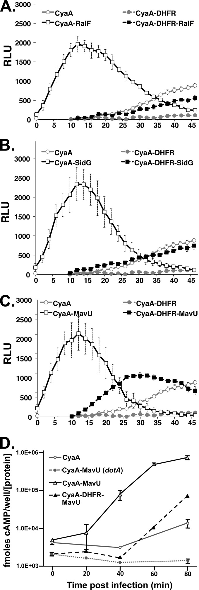
Fig 6.
The DHFR-IDTS fusion proteins exhibit kinetic defects in translocation. Results of real-time translocation assays of CyaA and Cya-DHFR moieties fused to IDTS RalF (A), SidG (B), and MavU (C) are shown. HEK293 cells transiently transfected with the pGloSensor cAMP biosensor plasmid and FcγRIIA were incubated with the pGloSensor luciferase substrate reagent for 1 h (see Materials and Methods). Cells were challenged with bacterial strains expressing the indicated proteins that had been opsonized with anti-Legionella IgG. The fluorescence was measured in a luminometer every 2 min for up to 1 h postinfection. Data represent the average of four independent replicates from a typical experiment ± standard error. (D) Direct quantitation of cAMP demonstrates kinetic defect of CyaA-DHFR-MavU fusion. CyaA translocation assays of CyaA-MavU and CyaA-DHFR-MavU fusion proteins were performed as described in the legend of Fig. 1 (see also Materials and Methods). Data represent the average of three replicates ± standard error from a typical experiment. RLU, relative light units.

