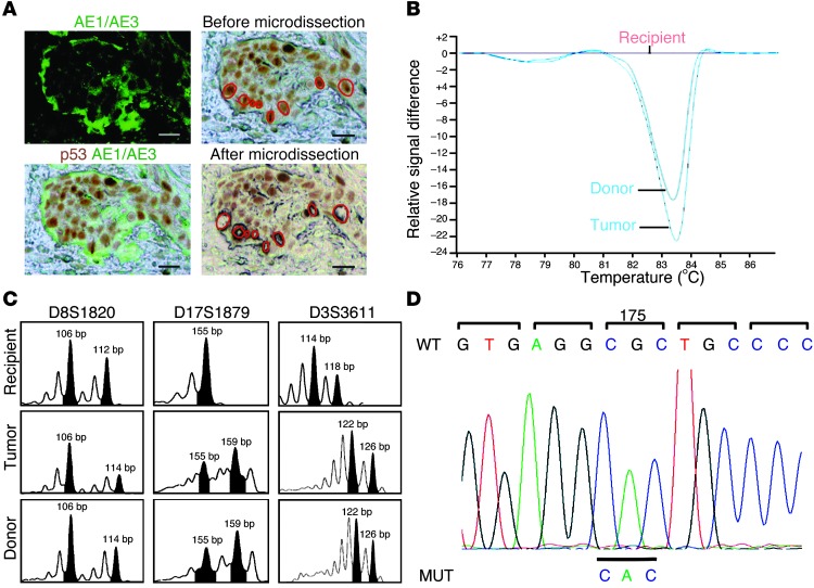
Figure 1. Recipient skin SCC.
(A) Laser microdissection of p53+AE1/AE3+ basal cells from invasive areas. Scale bars: 15 μm. (B) mtDNA-HRM of laser-microdissected p53+ tumor cells and donor and recipient normal cells. Laser-microdissected p53+ tumor cells and donor normal cells showed similar profiles. (C) DNA profiles from laser-microdissected p53+ tumor cells and donor and recipient normal cells. Microsatellite analysis at the D8S1820 locus showed the donor origin of the p53+ tumor cells (106 and 114 bp), similar to donor DNA, but different from recipient DNA (106 and 112 bp). At the D17S1879 locus, microdissected p53+ tumor cell and donor DNA were heterozygous (155 and 159 bp), but recipient DNA was homozygous (155 bp). (D) Sequencing of TP53 exon 5 PCR product identified a base substitution G>A on codon 175, corresponding to a R>H amino acid change in DNA, from laser-microdissected p53+ tumor cells.

