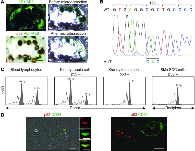
Figure 2. Donor kidney.
(A) Kidney graft biopsy performed 7 years before skin SCC diagnosis. Laser microdissection was used to select p53+AE1/AE3+ cells in renal tubules. Scale bars: 5 μm. (B) TP53 sequencing identified the same base substitution in laser-microdissected p53+ epithelial cells from the kidney graft tubules as in skin SCC tumor cells. (C) Since sequencing showed an homozygous mutation in both skin SCC and kidney graft tubules, we checked this result. Polymorphic microsatellite marker IGP53, located in intron 1, showed loss of heterozygosity for this locus in recipient skin SCC and p53+ kidney graft tubule cells, but not in donor blood lymphocytes or p53– kidney graft tubule cells. (D) Kidney graft biopsy with staining for p53 as well as the renal stem/progenitor cell marker CD24. Double-stained cells were few and were located in kidney tubules (arrow), not in the glomerular area (G). Scale bars: 25 μm; 10 μm (insets, enlarged ×3).

