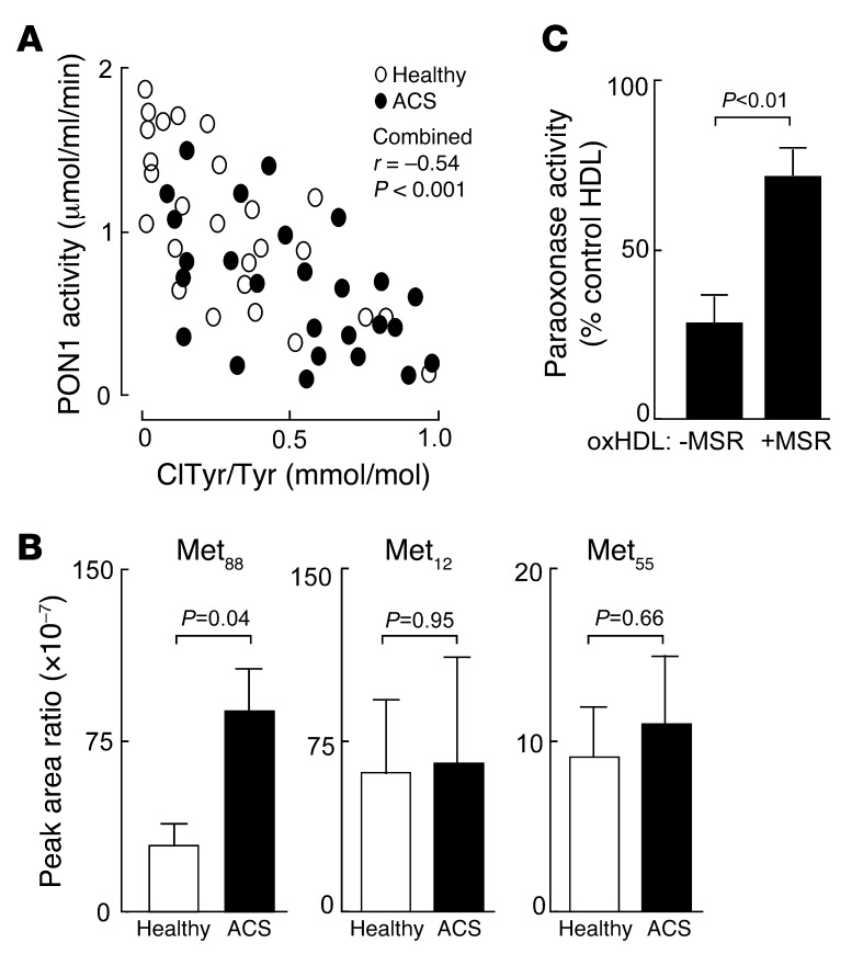
Figure 6. Methionine oxidation plays a role in PON1 activity and stability.
(A) Inverse correlation between systemic PON1 activity and protein content of ClTyr-isolated HDL from ACS (black ovals; n = 26) and healthy nondiabetic control subjects (white ovals; n = 26) from case-control cohort 1 (see Supplemental Table 1). P value shown is for the Spearman’s rank correlation between PON1 activity level and HDL ClTyr content among both control and ACS subjects. (B) Quantification of site-specific methionine oxidation (methionine sulfoxide) within PON1 recovered from isolated HDL from ACS (n = 10) and healthy nondiabetic control subjects (n = 10) enrolled in case-control cohort 2 (Supplemental Table 2). Results shown are expressed as the peak ratio of peptide harboring the indicated methionine sulfoxide residue or parent (methionine-harboring) peptide relative to the reference peptide, as described in Methods. (C) Isolated human HDL from a healthy donor was incubated with the MPO/H2O2/Cl– system (oxHDL), and then the effect of exposure to either methionine sulfoxide reductase (+MSR) or vehicle control (–MSR) on PON1 activity was determined as described in Methods. PON1 activity is expressed relative to paraoxonase activity measured in HDL prior to exposure to the MPO/H2O2/Cl– system. Data are the mean ± SD of triplicate determinations.

