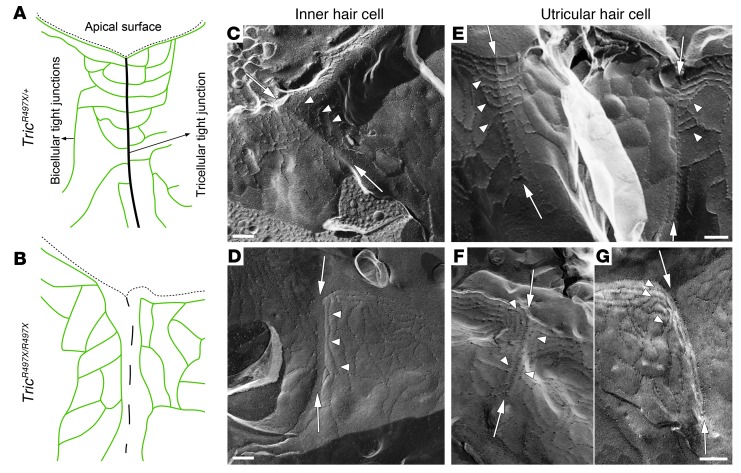
Figure 5. Tricellulin is required for the normal development of the tricellular junction structure.
(A and B) Schematic representations of the ultrastructure of tight junctions in the inner ear epithelia of TricR497X/+ and TricR497X/R497X mice. The thin dashed lines indicate the vertices of the apical surface. (A) In the controls, the bicellular tight junction strands (green) are associated with the tricellular tight junction (black line). (B) In the TricR497X/R497X mice, the tricellular tight junction is not complete (dashed line). Also, the topmost bicellular tight junction strands turn downward and run parallel to the tricellular tight junction. (C–G) Electron microscopy images of freeze-fracture replica of tricellular tight junctions in the (C and D) organ of Corti and (E–G) utricular macula of (C and E) TricR497X/+ and (D, F, and G) TricR497X/R497X mice. (C and E) In the TricR497X/+ animals, tricellular tight junctions appear as a “fishbone” (arrows), where the elements of the bicellular junctions meet the tricellular tight junction (arrowheads). (D, F, and G) In the TricR497X/R497X animals, the central element of the tricellular junction is irregular and appears to be formed of a chain of disconnected particles (arrows). The bicellular tight junction strands do not meet the tricellular junction (arrowheads); instead, they appear to turn around and join the neighboring bicellular junction strands. Scale bar: 200 nm (C–E; F and G).

