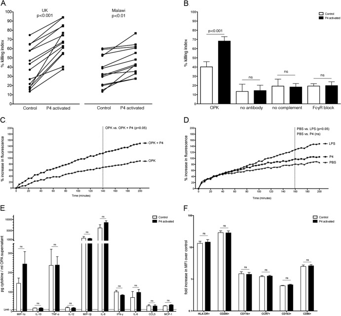
Fig 1.
(A) Opsonophagocytosis killing assay (OPKA) using human alveolar macrophages from healthy volunteers recruited in Liverpool, United Kingdom (n = 14), and Blantyre, Malawi (n = 13); (B) OPKA of United Kingdom volunteers (n = 6), where “OPK” designates assays in which all components are included (macrophage, bacteria, antibody, complement), “no antibody” designates assays without the presence of antibody, “no complement” designates assays without the presence of complement, and “FcγR block” designates assays where Fcγ receptors were occupied by IgG prior to the assay; (C) intracellular oxidation of alveolar macrophages (n = 6; United Kingdom population) during an opsonophagocytosis assay in the presence (OPKA+P4) or absence (OPK) of peptide stimulation; (D) intracellular oxidation in the presence of PBS, LPS (100 μg/ml), or P4 (1 mg/ml) alone; (E) cytokine levels detected in OPKA supernatant using a BD Flex Set kit following a 2-h OPKA; (F) surface markers of activation on control or P4-activated alveolar macrophages (n = 6) following a 2-h OPKA, stained and analyzed using flow cytometry.

