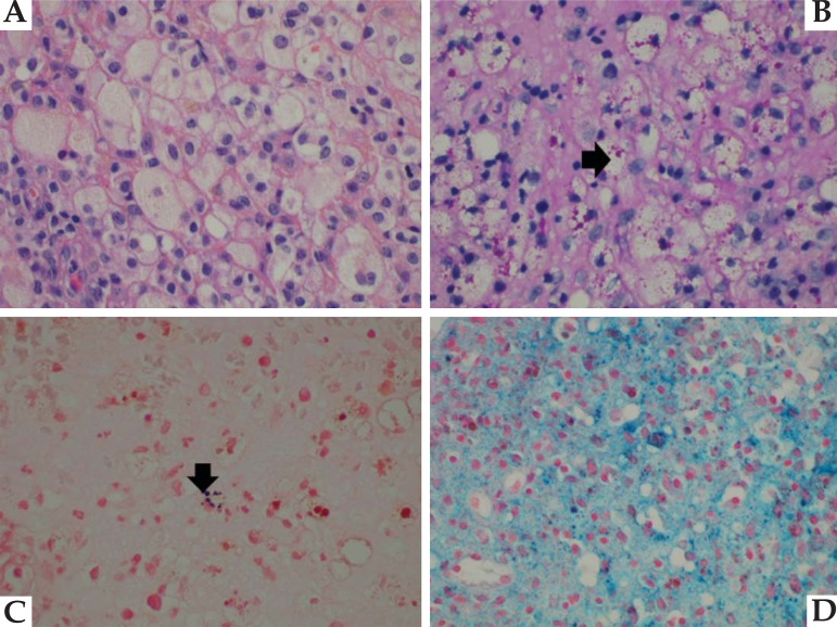
FIGURE 2.
Malakoplakia: A-hematoxylin-eosin (400 X) stain showing the sheets of macrophages. B-von Hansemann cells in PAS stain (400 X)(black arrow). C-Michaelis- Gutmann bodies, shown in black after Von Kossa staining (400 X)(black arrow). D-Prussian blue demonstrates hemosiderin inside macrophages (400 X)

