FIGURE 2.
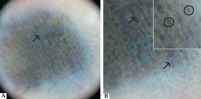
(a) Dermoscopy (10x): area composed of blue-grey structures across a yellow background. In the middle of the lesion there was an area with a radiated aspect corresponded to scar fibrosis caused by the earring. (b) Dermoscopy (40x): three types of dermoscopic structures existed: annular structures (black circles), short linear structures (arrows) and multiple spots (blue circle)

