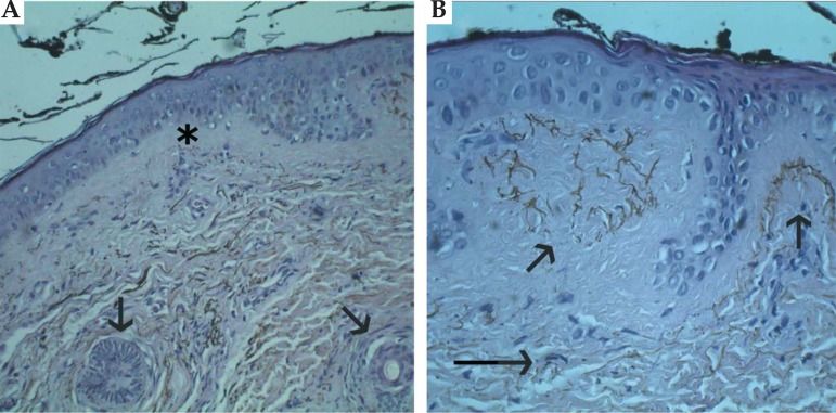
FIGURE 3.
(a) Histopathology(100x): brown-pigmented granules surrounding the eccrine glands (arrows). The pigment was localized under a Grenz zone (asterisk) (b) Histopathology (200x): brown-pigmented granules were inside the dermic papillae (short arrows) and densely populated the interpapillary dermis over the elastic fibres and vessel walls (long arrows)

