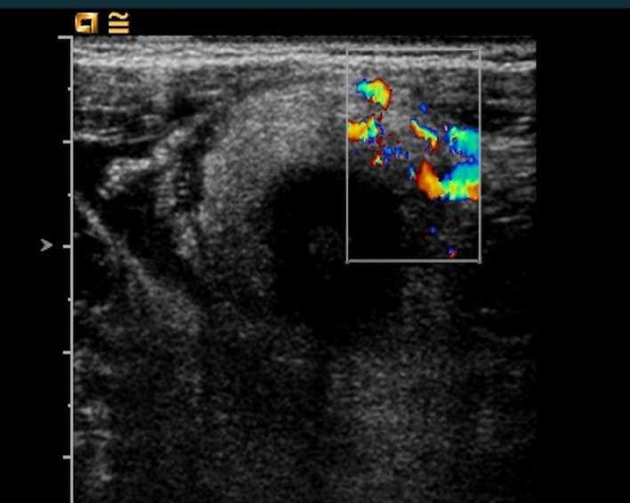
Figure 1:Abdominal ultrasound image of ileocecal wall thickening (3-6 mm) with increased signal in the vascular color-doppler and hyperplasia of the surrounding fatty tissue.

Figure 1:Abdominal ultrasound image of ileocecal wall thickening (3-6 mm) with increased signal in the vascular color-doppler and hyperplasia of the surrounding fatty tissue.