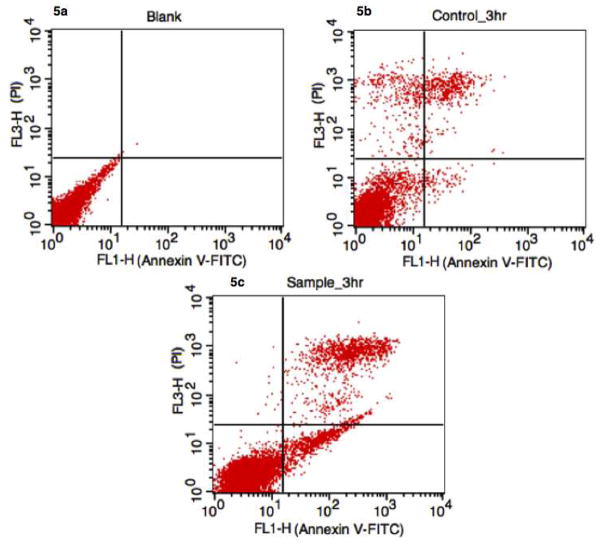
Figure 4.
Flow cytometry analysis of anti-CD95 induced apoptosis using Annexin V-FITC/PI. Apoptotic cells are located in the lower right quantrant. (a) Control (probe without anti-CD95) and (b) blank (no probe or antibody) showed normal levels of nectrotic and apoptotic cells. However, when receptor mediated apoptosis was initiated, apoptotic cells were observed at 3 hours (c). Given the rapid temporal resolution of our mitochondrial-based assay, it is possible to exclude mitochondrial protein release as a co-mechanism during receptor-mediated apoptosis in Ramos cells.

