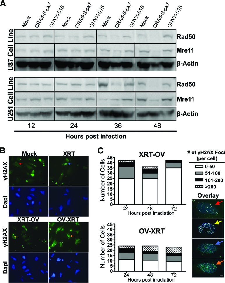
Figure 7.
Radiosensitizing effect of CRAd-Survivin-pk7 (CRAd-S-pk7) infection on glioma. (A): Protein expression of the Mre11-Rad50-NBS1 complex proteins Rad50 (153 kDa) and Mre11 (81 kDa) at 12, 24, 36, and 48 hours after infection with 50 infectious units of CRAd-S-pk7 or ONYX-015. Western blots show that Rad50 and Mre11 protein expression are reduced at both 36 and 48 hours after infection with CRAd-S-pk7 but not ONYX-015. (B): Immunofluorescent staining of radiation induced γH2AX foci under a confocal laser microscope. Top: Anti-γH2AX (green); bottom: Dapi (blue). Magnification, ×63. Scale bar = 20 μm. (C): Quantification of γH2AX foci resolution over 72 hours after XRT treatment. The number of γH2AX foci per cell was counted and grouped according to the following range of foci per cell: 0–50 (red arrows), 51–100 (yellow arrows), 101–200 (blue arrows), and >200 (orange arrows). Left: Time effect was determined by ordinal logistic regression analysis. The number of γH2AX foci was significantly resolved over time in XRT-OV-treated cells (p = .020), whereas there was no significant change in the number of foci over time in OV-XRT-treated cells (p = .386). Right: Representative overlay images of each range of foci per cell (anti-γH2AX, green, and anti-DAPI, blue). Magnification, ×63. Scale bar = 20 μm. Abbreviations: Dapi, 4′,6-diamidino-2-phenylindole; OV-XRT, radiation therapy 24 hours after oncolytic virus; XRT-OV, oncolytic virus 24 hours after radiation therapy.

