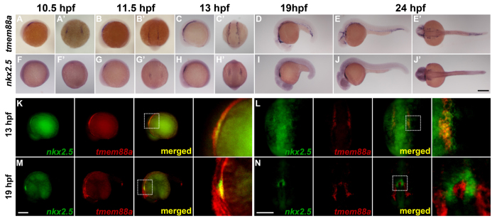
Fig. 2.

In situ hybridization of tmem88a and nkx2.5 in developing zebrafish embryos. (A-J′) tmem88a and nkx2.5 expression during early development in zebrafish. Whole-mount in situ hybridizations for tmem88a (A-E′) and nkx2.5 (F-J′) were performed on embryos ranging from 10.5 hpf to 24 hpf. (K-N) Double fluorescent in situ hybridizations for nkx2.5 (green) and tmem88a (red) at 13 hpf (eight-somite stage; K,L) and 19 hpf (M,N). The white dotted box in merged panels (K-M) outlines the region shown at 4× magnification to the right. A-J, K and M are lateral views with anterior to the left. A′-J′, L and N are dorsal views with anterior to the bottom. Scale bars: in J′, 200 μm for A-J′; in M, 200 μm for K,M; in N, 200 μm for L,N.
