
Fig. 5.
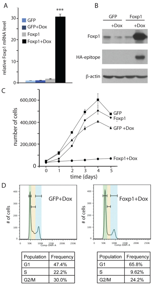
Overexpression of Foxp1 leads to cell cycle arrest. (A) Real-time PCR analysis shows Foxp1 is induced in tet-regulatable manner in tet-inducible Foxp1 keratinocytes. Keratinocytes were transduced to express tet-inducible HA-tagged Foxp1 or GFP and were selected with G418. Real-time PCR was performed on cDNA synthesized from RNA that was isolated from the transduced cells that were grown in media with doxycycline (Dox) or vehicle for 24 hours. Data are mean ± s.e.m. ***P<0.001. (B) Western analysis verifies that tet-inducible HA-Foxp1 was induced in a Dox-dependent manner. Keratinocytes were transduced to express tet-inducible HA-tagged Foxp1 or GFP and were selected with G418. With antibodies against Foxp1 or HA-epitope, western analysis was performed on protein lysates that were isolated from the transduced cells that were grown in media with Dox or vehicle for 24 hours. (C) Growth curve demonstrates that expression of Foxp1 results in a cessation of growth. Twenty thousand tet-inducible Foxp1- or GFP-expressing cells were plated onto each well of a 24-well plate. A day later, cells were counted as t=0 and Dox or vehicle were added to the cells. Cells were counted in duplicate every 24 hours subsequently. Error bars represent s.d. (D) Cell cycle profile of cells induced to express Foxp1 shows that overexpression of Foxp1 results in an increased number of cells in the G1 phase. Cell cycle analysis was performed on keratinocytes expressing tet-inducible Foxp1 or GFP that were grown with Dox or vehicle for 24 hours. Green, yellow and blue bands represent G1, S and G2/M, respectively.