Fig 1.

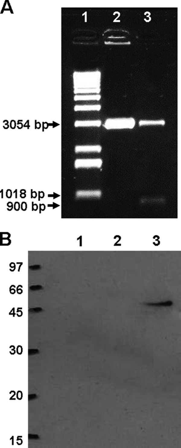
Plasmid construction and expression of Apa antigen. (A) Amplified fragments corresponding to kDNA of pVAXapa plasmid construction. The amplified products were separated by electrophoresis on a 2.5% agarose gel and stained with ethidium bromide. The image of the gel captured under UV light shows the 900-bp fragment corresponding to apa and the 3,054-bp fragment corresponding to the pVAX plasmid. Lane 1, molecular size standard (1 kb of DNA); lane 2: pVAX digested with XhoI; lane 3: pVAX-apa digested with XhoI and EcoRI. (B) Transient expression of Apa by pVAX-apa: Western blot analysis of cell lysates from BHK-21 cells transfected with pVAX-apa (lane 3) or pVAX (lane 2) or not transfected (lane 1). The size of the molecular mass standard is indicated, in kilodaltons. The primary antibody used was rabbit anti-Apa serum.
