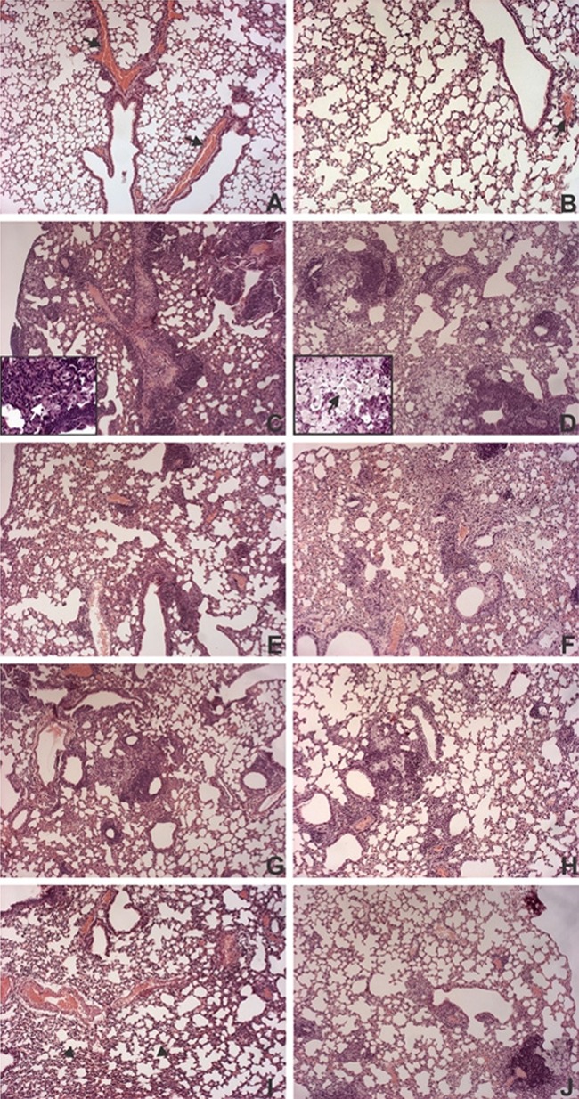
Fig 3.
Histopathology of lungs from immunized mice at 30 and 70 days after M. tuberculosis infection. BALB/c mice (n = 5) were immunized s.c. with a single dose of BCG (5 × 10⁵ CFU; BCG group) or one dose of BCG administered s.c., followed by one dose of pVAX-TDM-Me (BCG-PVAX) or pVAXapa-TDM-Me (BCG-APA) administered i.m. after 30 days. Thirty days after vaccination, the mice were challenged intratracheally with a virulent strain of M. tuberculosis (1 × 10⁵ CFU). At 30 (A) or 70 (B) days after infection, the lungs were fixed in buffered formalin and stained with hematoxylin and eosin. Representative images of histological sections of lung lobes are shown for the PBS (A and B), TB (C and D), BCG (E and F), BCG-PVAX (G and H), and BCG-APA (I and J) groups. White and black arrows in the insets (TB group) indicate giant cells and xanthomatous macrophages, respectively. Arrows indicate congestion of blood vessels, and arrowheads indicate collapsed pulmonary alveoli. Original magnification, ×100. Magnification of the insets, ×400. The results shown are representative of 3 independent experiments.

