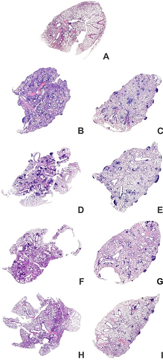
Fig 4.

Overview perspective of the whole lungs of mice at 30 and 70 days after M. tuberculosis infection. BALB/c mice were immunized as described in the legend to Fig. 3. The lungs were fixed in buffered formalin and stained with hematoxylin and eosin. Representative images of lung lobes are shown for the PBS (A), TB (B and C), BCG (D and E), BCG-PVAX (F and G), and BCG-APA (H and I) groups. The results shown are representative of 3 independent experiments.
