Fig 3.

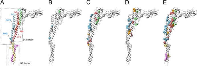
Locations of the first mutation and second mutation sites on the ribbon model of the R-type flagellin. (A) The helices 1, 2, 3, 4, and 5 are represented by violet, light coral, pale green, sky blue, and khaki, respectively. The helices 1and 5 and 2, 3, and 4 are categorized into D0 and D1 domains, respectively. The first mutation sites Gly426, Ala449, Gly70, and Ala80 are represented using a CPK model. Gly426 and Ala449 are sky blue, and Gly70 and Ala80 are light coral. (B to E) The second mutation sites of SJW1655, SJW1660, HFG180, and HFG195 revertants, respectively. The second mutation sites are represented by a CPK model. The second mutation sites located on the helices 1, 2, 3, 4, and 5 are violet, light coral, pale green, sky blue, and khaki, respectively. The residues not located on helices 1 to 5 are orange.
