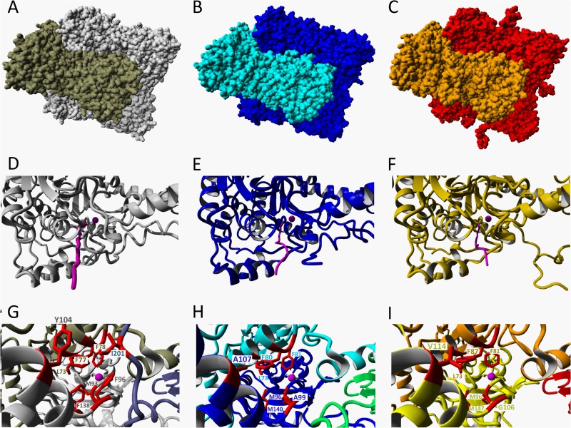
Fig 7.
Crystal structure and models of CS2 hydrolases. (A) Resolved crystal structure of the CS2 hydrolase from Acidianus strain A1-3 and structural models of the CS2 hydrolases from A. thiooxidans strain G8 (B) and strain S1p (C), modeled using the hexadecameric structure of the Acidianus A1-3 CS2 hydrolase as a template. The two octameric interlocking rings forming the hexadecamer are shown in different colors. (D, E, F) Details of octamers showing the narrow hydrophobic entrance tunnels (pink) to the Zn-containing active site in the Acidianus CS2 hydrolase (D) and in the models of the G8 (E) and S1p (F) CS2 hydrolases. The Zn atoms are indicated by pink spheres. (G, H, I) Views of the tunnels looking in from the entrances to the active sites, showing the three monomers (two shades of gray and blue) and the residues (red) forming the tunnel in the Acidianus CS2 hydrolase (G) and in the models of the G8 hydrolases (blue, cyan, and green monomers) (H) and the S1p CS2 hydrolases (yellow, orange, and red monomers) (I).

