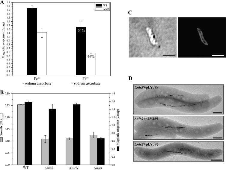
Fig 6.
(A) Magnetic response (Cmag) of WT and ΔnirS mutant after three serial transfers under microaerobic conditions in iron-deprived medium. Nonmagnetic WT and the ΔnirS mutant were induced anaerobically for 6 h in the presence of reduced (100 μM ferrous chloride plus 0.2 mM ascorbate) and oxidized (100 μM ferric chloride) iron sources. (B) Growth (based on the OD565) and magnetic response (Cmag) of WT, ΔnirS, ΔnirN, and Δnap strains in an incubator hood with a constant atmosphere of 2% O2 and 98% N2. (C) Intracellular localization of the MSR-1 NirS protein N-terminally tagged with mCherry in the ΔnirS mutant. Differential interference contrast microscopy (left) and fluorescence microscopy (right) were used. Bar, 2 μm. (D) TEM images of the anaerobically grown ΔnirS mutant carrying nirS (top to bottom) from M. magneticum (amb1395, amb4165) and P. stutzeri (PST_3532), respectively. Bar, 500 nm.

