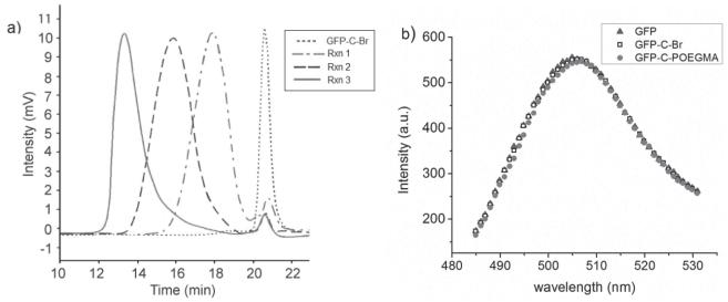
Figure 2.
a) Size exclusion chromatography (SEC) traces of GFP-Br (dotted line), Rxn 1 (dashed dotted line), Rxn 2 (dashed line), and Rxn 3 (solid line). b) Fluorescence spectra of GFP before initiator attachment (solid triangle), after initiator attachment (empty square), and after in situ ATRP (solid circle, ATRP at Rxn 3 conditions: 263.1 kDa as measured by SEC-MALS); all samples at 20 μM.

