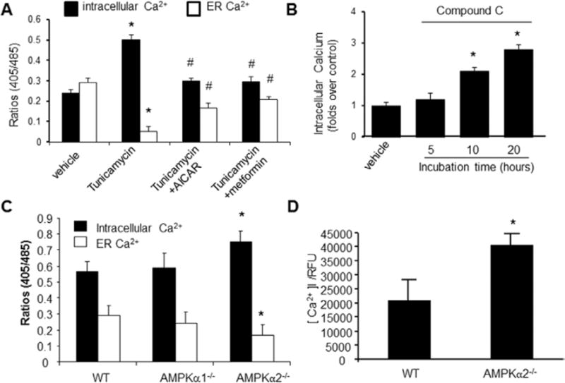
Figure 4.

AMP-activated protein kinase (AMPK) activation on intracellular Ca2+ and endoplasmic reticulum (ER) Ca2+ in cultured vascular smooth muscle cells (VSMCs). After being loaded with Indo-1/AM dye, the cells were stimulated with ionomycin (10 μmol/L), which triggers Ca2+ release from the ER. A, 5-Aminoimidazole-4-carboxamide ribonucleotide (AICAR) and metformin suppress the rise of intracellular [Ca2+]i, whereas they increase Ca2+ in the ER. *P<0.05 Tunicamycin vs vehicle; #P<0.05 AICAR or metformin plus Tunicamycin vs Tunicamycin alone; n=5. *P<0.05 Tunicamycin vs vehicle; #P<0.05 Tunicamycin vs Tunicamycin plus AICAR or metformin. B, Effects of compound C on ionomycin-induced Ca2+ release and intracellular Ca2+ store; *P<0.05 compound C vs vehicle; n=6, *P<0.05 compound C vs vehicle. C, Increased levels of intracellular Ca2+ and decreased Ca2+ in ER in VSMCs derived from AMPKα2−/−; AMPKα2−/− vs wild-type (WT); n=7; *P<0.05 WT vs AMPKα2−/−. D, Increased [Ca2+]i/relative fluorescence units (RFUs) in VSMCs from AMPKα2−/− mice. AMPKα2−/− vs WT. n=8. Statistical analysis was performed by using a 2-tailed Student t test between 2 groups.
