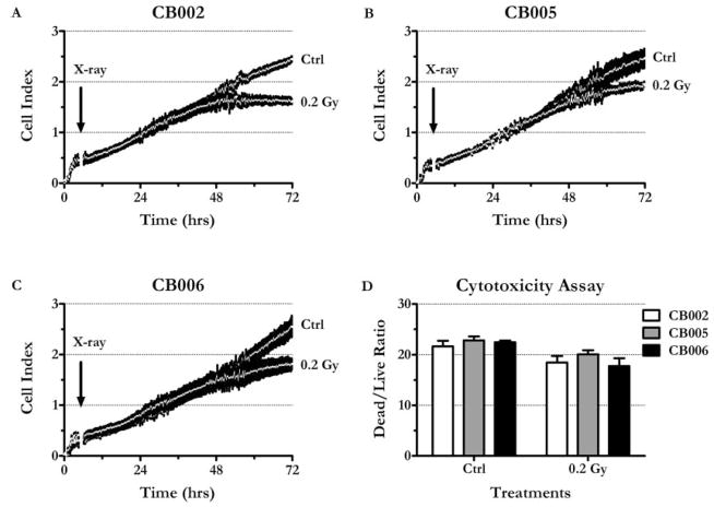
Figure 2.
LDIR inhibits ECFC growth. A, CB002. B, CB005. C, CB006. Cells were seeded at 2,000 cells per well in 96-well E-plates and were growing for 72 hr. Impedance (expressed as nondimensional “Cell Index”) was measured every 15 min. Arrow indicates the time of irradiation (0.2 Gy X-rays, ~ 4.5 hr after the seeding). Graphs were generated by plotting Cell Index mean values ± SEM vs. time (n = 4). Note that means form a continuous white line. D, MultiTox-Fluor Assay demonstrates that radiation does not increase dead/live cell ratio. CB002, CB005, and CB006 were seeded in black-walled clear plates, irradiated, and incubated as described elsewhere. At 72 hr, fluorescent protease substrates (Promega, Madison, WI) were added directly to the media, and the reaction was incubated for 2 hr. 400ex/505em and 485ex/520em filters were used to detect the products of live and dead cell-associated proteases, respectively. Mean values ± SEM are shown (n = 4).

