Figure 8.
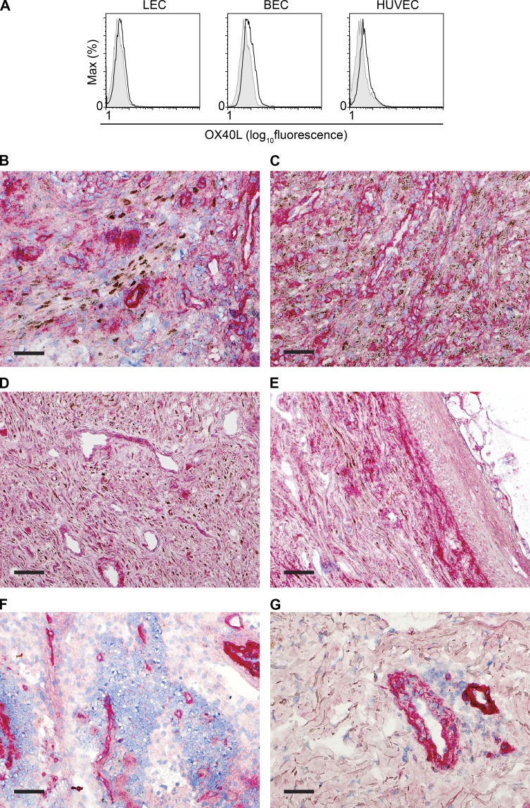
OX40L is abundantly expressed in KS lesions. (A) Expression of OX40L on the cell surface was assessed with flow cytometry in three types of primary endothelial cells: human dermal microvascular lymphatic endothelial cells (LEC), human dermal microvascular blood endothelial cells (BEC), and HUVECs. One result representative of two independent experiments is shown. (B–G) OX40L (red) and HHV-8 LANA (brown) expression was assessed by immunohistochemistry in frozen tissue sections. Bars, 5 mm. (B–E) AIDS-related KS lesions in lymph node (B), presenting as a submental mass (C), and in skin (D and E) show HHV-8–positive cells (punctate nuclear brown staining) as well as numerous OX40L-positive cells. (F and G) Non-KS tissues used as controls include normal lymph node (F) and skin with mycosis fungoides (G), showing OX40L in cells lining vascular spaces.

