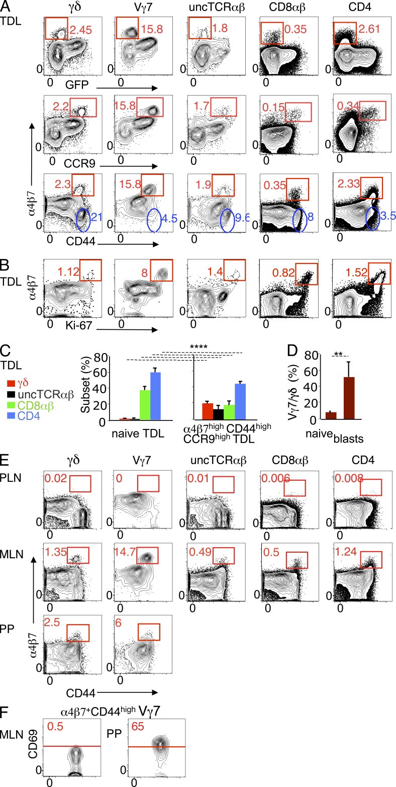
Figure 3.
Conventional and unconventional cycling TDL blasts express the highest levels of gut-tropic molecules and are GALT-related. TDLs of 8–12-wk-old RAG2p-GFP mice were assessed for expression of gut-tropic molecules by flow cytometry. (A) Dot plots show α4β7 versus GFP, CCR9, or CD44 (eight mice individually studied in seven independent experiments). Red gates define cells expressing high levels of α4β7 lacking GFP or coexpressing high levels of CD44 or CCR9. α4β7 MFI in α4β7high GFP− (CD44high) versus GFP+ (CD44low) TDLs: γδ, 15133/1758; Vγ7, 15139/1833; uncTCRαβ, 15321/780; CD8αβ, 7427/369; CD4, 5113/397. Blue gates indicate CD44high TDLs that lack α4β7. (B) Dot plots showing α4β7 versus Ki-67 expression (three mice in three independent experiments). (C) T cell subset distribution in naive (CD44low) and α4β7high CCR9high CD44high TDLs from the mice described in A. ****, P < 0.0001. (D) Frequency of Vγ7+ cells in naive (CD44low) and α4β7high CCR9high γδ TDLs (four mice in four independent experiments). **, P < 0.003. (E). Dot plots showing CD44 versus α4β7 expression in MLN, PLN (pool of brachial, axillary and inguinal LN) and PPs. MLN and PPs data are representative of 6 independent experiments, each with a pool of 2–3 mice. Two of these experiments included PLN, which in each case were a pool from 3 mice. (F) CD69 expression of α4β7high CD44high Vγ7+ cells in MLN and PPs, as gated in the corresponding panels in E.

