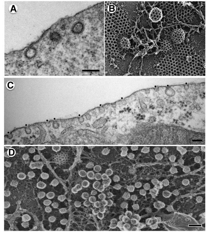
Figure 1. Thin section and quick-freeze deep etch images of clathrin coated vesicles and caveolae.
(A) Thin section, TEMs reveal the bristled appearance of clathrin-coated pits in fixed and plastic embedded mouse fibroblasts (from [27]), bar = 200 nm. (B) Quick-freeze deep-etch images reveal the lattice-like coat on flat and rounded clathrin coated pits, courtesy of John Heuser. (C) Ultra thin section EM of caveolae in fixed and plastic embedded 3T3-L1 adipocytes (from [18]). Bar = 100 nm. (D) Deep-etch EM replica of differentiated 3T3-L1 adipocyte produced in the Heuser lab by N. Morone, iCeMS, Kyoto University, Japan. Bar = 100 nm.

