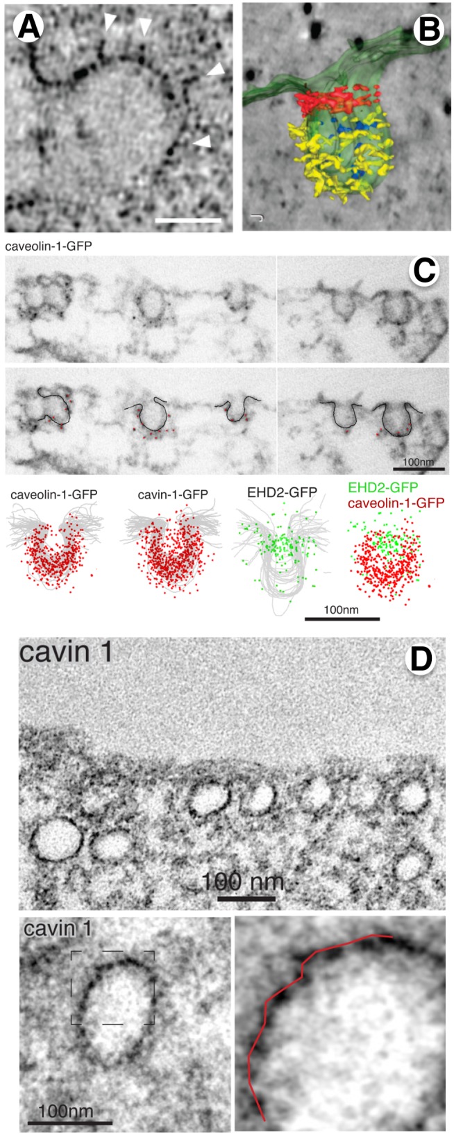
Figure 2. Electron microscopy tomography, immunolocalization, and the use of genetically encoded electron microscopy markers reveal high resolution ultrastructure of caveolae.

(A) Thin section electron micrograph of a single caveolae in 3T3-L1 adipocyte prepared by high pressure-freezing/freeze-substitution reveals an irregular coat. Bar = 30 nm (from [18]). (B) Tomographic reconstruction of caveolae from 3T3-L1 adipocytes reveals electron dense material around the bud and neck (from [18]). (C) Pre-embedding labeling with nano-gold secondary antibodies reveals differential localization of caveolae coat components (CAV1, cavin-1) on the buds, relative to the EHD localization at the neck (from [20]). (D) Cavin-1-miniSOG fusion protein stably expressed in RPE cells allows localization of the cavin-1 protein by EM. The ∼10 nm periodicity of the DAB reaction product can be seen in the higher magnification images (from [20]).
