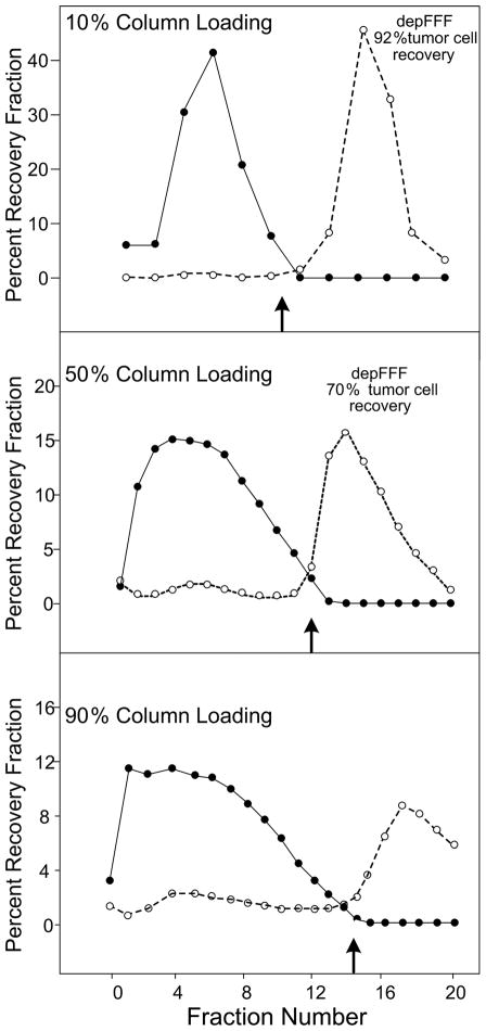
Figure 3.
Elution profiles of PBMN (●) and cancer cells (❍) as a function of the percentage of the depFFF volume that was loaded with sample prior to cell settling. The DEP frequency was switched from 60 kHz to 15 kHz at the fractions shown by the small arrows. Better recovery of tumor cells occurred if the cells remaining in the depFFF were flushed rapidly from the chamber after the DEP frequency was changed to 15 kHz (Flush recovery).

