
Figure 7. Tumor MMP-1 functionally regulates the permeability of intratumoral vasculature via PAR1-mediated signaling.
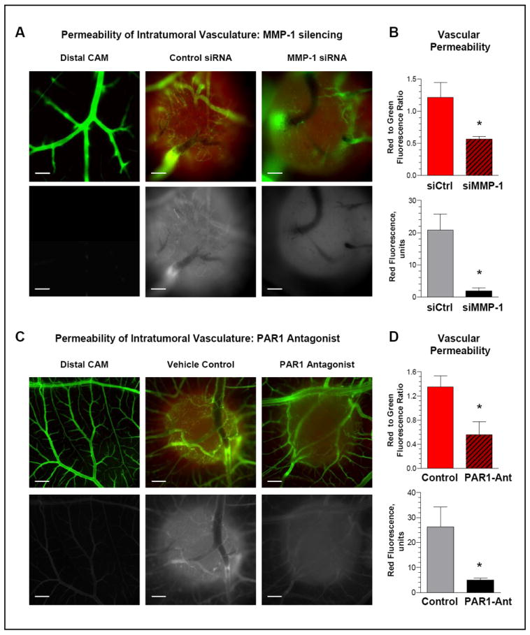
(A) Permeability of intratumoral vasculature is regulated by tumor-produced MMP1. Topical CAM microtumors were initiated from non-labeled HEp3-hi/diss cells. Six days after cell grafting, the embryos were injected i.v. with the permeable low mol. wt. TRITC-dextran. After 1-hr incubation, the embryos were inoculated with the non-permeable high mol. wt. FITC-dextran. The portions of the CAM with and without microtumors were visualized in fluorescent microscope and images were acquired at an original 10x magnification. Top panels, green and red fluorescence signals are merged. Bottom panels, red fluorescence is depicted monochromatically in white in order to highlight differential exudation of low mol. wt. dextran. Bar, 200 μm.
(B) Quantitative analysis of intratumoral permeability. Following imaging, individual microtumors were lysed and levels of red and green fluorescence were measured at 557 and 492 nm, respectively. Top graph, Permeability of intratumoral vasculature in individual tumors is presented as ratio of dextran exudation (red fluorescence) to total volume of perfusable vasculature (green fluorescence). Bottom graph, levels of dextran exudation presented as red fluorescence units. Data are means±SEM; *, P<0.05.
(C) Permeability of intratumoral vasculature is regulated by PAR1-mediated signaling. Developing HEp3-hi/diss microtumors were treated on day 2 and 4 with the PAR1 antagonist SC79797 (PAR1-Ant) or vehicle control. Dextran permeability in the distal CAM and microtumors was analyzed as described in (A). Original magnification, 4x. Bar, 500 μm.
(D) Quantitative analysis of intratumoral permeability. CAM microtumors treated with PAR1 antagonist SC79797 (PAR1-Ant) or vehicle (Control) were dissected and lysed. Vascular permeability was measured as described in (B). Data are means±SEM; *, P<0.05.