
Figure 5.
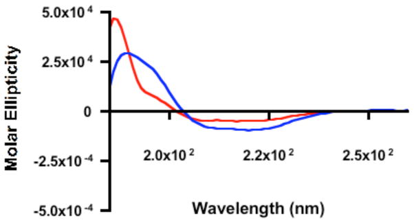
Circular dichroism of RSV F protein (red trace) or RSV F protein-nanorod conjugates (blue trace) suggests an intact secondary structure after F protein-nanorod conjugation. Specifically, F protein: helix: 22%, beta sheet: 42% random coil: 36%; F protein-nanorod conjugates: helix: 35%, beta sheet: 31% random coil: 34%.