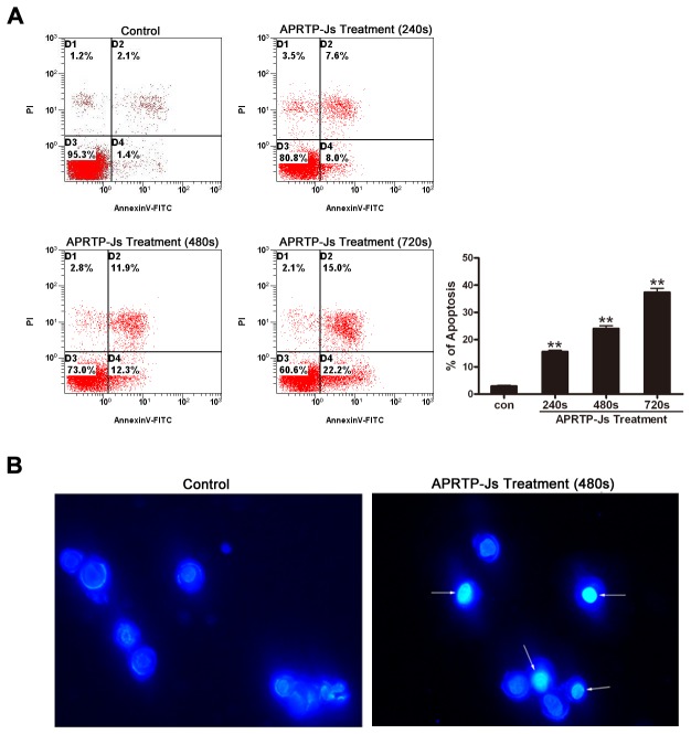
Figure 3. APRTP-Js treatment induced apoptosis in HepG2 cells.
Cells were treated by APRTP-Js for 240 s, 480 s and 720 s, and then cultured for 24 h. (A) Cells were double stained with annexin V-FITC and PI and analyzed by flow cytometry. Cells that stained positive for annexin V-FITC and negative for PI were undergoing early stage of apoptosis; Cells that stained positive for both annexin V-FITC and PI were in the end stage of apoptosis; Cells that stained negative for both annexin V-FITC and PI were alive and not undergoing measurable apoptosis. Percentage of apoptotic cells (annexin V-FITC positive) was shown by histogram. (B) Observation of Hoechst 33342 apoptosis staining by fluorescence microscopy. Examples of typical apoptotic nuclei were indicated by white arrows.

