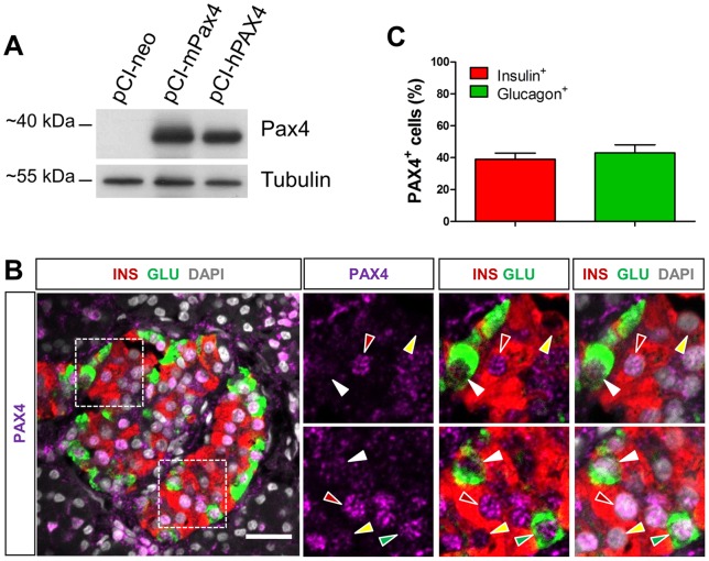
Figure 1. PAX4 is detected in both human pancreatic alpha and beta cells.
(A) Detection of ectopically expressed mouse and human PAX4 by western blotting. The capacity of the anti-Pax4 antibody to recognize human PAX4 was assessed using αTC1-9 cells transfected respectively with the pCI-neo empty vector or constructs expressing mouse Pax4 (mPax4) or human PAX4 (hPAX4) protein by western-blotting analysis. Protein extracts from transfected cells were used for the detection, using antibody against Pax4. (B) Representative images of triple IF staining with antibodies against PAX4 together with glucagon and insulin. Right panels are the amplified view of the insets in the left panel. Red arrowheads, PAX4+ INS+ cells. Yellow arrowheads, PAX4−INS+ cells, Green arrowheads, PAX4+GLU+. White arrowheads, PAX4−GLU+ cells. (C) Percentages of insulin+ cells and glucagon+ cells expressing PAX4 represented as the averaged counting results ±S.E.M from n = 5 control individuals (A total of 164 GLU+ cells and 904 INS+ cells were analyzed). Scale bars = 25 µm.

