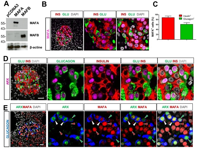
Figure 2. MAFA is detected in both human pancreatic alpha and beta cells.
(A) Detection of ectopically expressed human MAFA and MAFB by western blotting. The specificity of selected antibodies against human MAFA and MAFB were evaluated using mouse embryonic fibroblasts (MEF), which do not express endogenous murine MafA and MafB proteins, transfected respectively with constructs expressing human MAFA or MAFB protein by western-blotting analysis. Protein extracts from MEF transfected with empty pcDNA and pcDNA expressing respectively human MAFA and MAFB were used for the detection, using antibodies against MAFA (Abcam) or MAFB (anti hMAFB2, mouse monoclonal antibodies, clone 1F4). Note that the anti-MAFA antibody and the noncommercial anti-hMAFB2 reacted specifically without cross-reaction, whereas the other tested commercially available anti-MAFB antibodies failed (data not shown) (B) Triple immunofluorescent (IF) staining showing MAFA expression in human islets from healthy donors. (C) The percentages of cells expressing MAFA were 88.3±6.3% MAFA+ beta cells and 61.2±6.4% MAFA+ alpha cells. Results are the averaged expression ±S.E.M of counting results from n = 4 control individuals (1058 INS+ cells and 345 GLU+ cells were counted in total). (D) ARX expression was detected in human pancreatic alpha cells but not human pancreatic beta cells. Representative images of triple IF-staining showing ARX expression in human islets from healthy donors. ARX was detected only in nuclei of alpha cells. (E) Co-localisation of MAFA with ARX in human islets. Right panels are the amplified view of the inset in the left panel. Red arrowheads, MAFA+ARX−GLU−cells. Green arrowheads, MAFA−ARX+GLU+. White arrowheads, MAFA+ARX+GLU+ cells. Scale bar = 25 µM.

