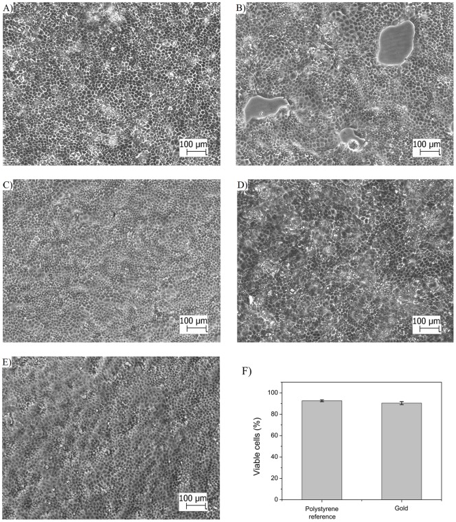
Figure 2. MDCKII cell morphology and viability on polystyrene and gold coated SPR sensor slide.
Light microscopy images of MDCKII cells cultured on A) polystyrene with a cell seeding density of 1×10⁵ cells/cm2, B) SPR sensor slide with a cell seeding density of 5×10⁴ cells/cm2 , C) SPR sensor slide with a cell seeding density of 7×10⁴ cells/cm2 , D) SPR sensor slide with a cell seeding density of 1×10⁵ cells/cm2, E) SPR sensor slide with a cell seeding density of 7×10⁴ cells/cm2 after exposing the cell monolayer to increasing concentration (2.5 μM, 25 µM and 250 µM) of propranolol during 1 hour at a flow rate of 10 μl/min in the SPR flow channel, F) cell viability of MDCKII cells grown on polystyrene reference and gold coated SPR sensor slides. The cell seeding time used for the cell monolayers in A)–F) was 3 days. The scale bar in all images is 100 µm.

