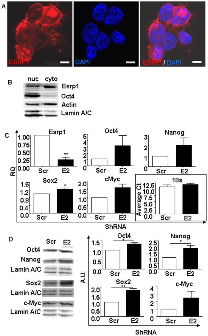
Figure 2. Pluripotency-related genes expression is affected by ESRP1 depletion.
A. Confocal microscopy analysis of ESRP1 (red) in mouse ES cells reveal that the protein is expressed not only in the nucleus by also in the cytoplasm of these cells. Nuclei are stained with DAPI (blue). Scale bar is 5 µm. B. Representative Western blot analysis shows that ESRP1 is expressed mainly in the cytoplasm of mouse ES cells. As expected, Oct4 has a main nuclear localisation. Actin and Lamin A/C were used for normalisation. C. qRT-PCR analysis of Esrp1, Oct4, Nanog, Sox2, and c-Myc mRNA in Scr and Esrp1-depleted ES cells shows that there was a increase in their expression. RQ is relative quantity (n = 4). D. Representative Western blot analysis of Oct4, Nanog, Sox2 and c-Myc expression in nuclear extracts from Scr and Esrp1-depleted ES cells. Densitometric analysis of the Western blots is shown; A.U. is arbitrary unit (n = 3).

