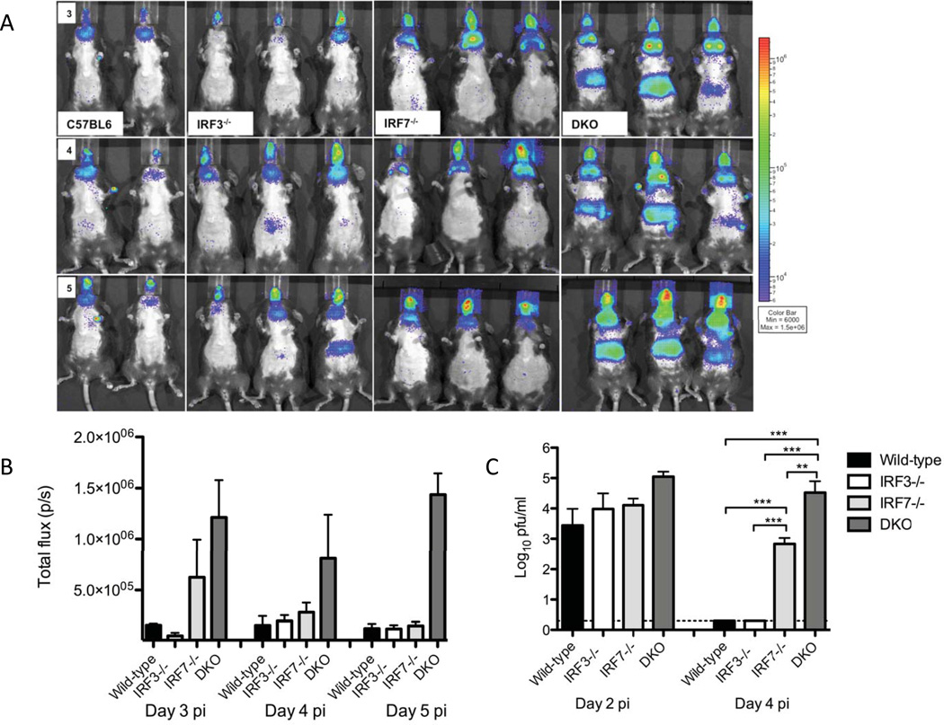
Figure 1. Pattern of viral spread in wild-type, IRF-3−/−, IRF-7−/− and DKO mice.
(A) In vivo bioluminescent imaging analysis was performed on days 2 through 5 post corneal infection with McKrae/DLux. Daily images for the same mice are shown on identical photon flux scales and are representative of 2 independent experiments. (B) Region of interest analysis of lymph node bioluminescence. ROIs were drawn around the lymph node area using the Living Image and IgorPro software and bioluminescent signal was reported in photons/sec. (C) Viral titers in the blood stream. Serum was isolated from mice two and four days post infection with McKrae/DLux and assayed on vero cell monolayers (n=4 mice per group, 2 independent experiments).

