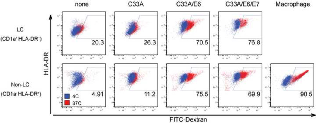
Figure 4. Increased phagocytic capacity of monocytes differentiated in the presence of HPV16 E6-transduced cell line.
CD14+ monocytes were co-incubated with UV-irradiated HPV-negative (C33A), HPV16 E6 C33A cells, or HPV16 E6 + E7 C33A cells in the presence of GM-CSF/ IL-4/TGF-β (LC condition) or GM-CSF (Macrophage condition) for 7 days. (A) Differentiated monocytes were incubated with FITC-Dextran for 1 hour at 37°C or 4°C. Phagocytic activity of CD1a+HLA-DR+ (upper row) and CD1a− HLADR+ (bottom row) populations at 37°C (red dots) and 4°C (blue dots) was analyzed by flow cytometry. These data were representative of three separate experiments.

