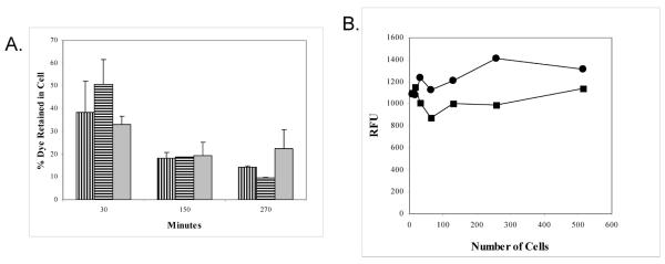
Figure 4.

A. Level of dye retention of Cell Tracker ( ), Vybrant (
 ), and Calcein-AM (
 ) labeled Jurkat cells over time.
B. Level of dye retention of Jurkat cells labeled with 2 μM (■) or 6 μM (●) concentrations of PKH67. Two fold dilutions of cells were performed and fluorescence was measured in a FL600 at 485ex/530em to determine the sensitivity of the signal.
