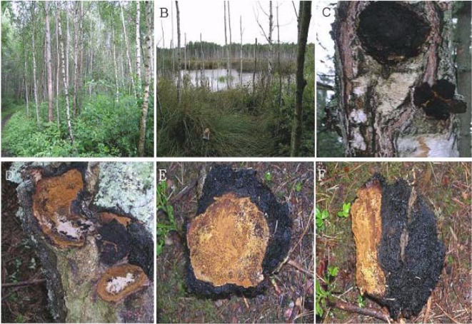
Fig. 1.
Plantation area of birch and sterile conk development of Chaga. A, Location of birch stand area in peat bog site; B, Wetland of birch stand in peat bog; C, Infected tree by I. obliquus. The black conk is the mycelia mass of the fungus; D, Infested area visualized by cutting off the cinder conk. The scar of the infected portion is evident as a yellowish-brown color; E, The front view of a sterile conk cut off from a host plant. The yellowish-brown color is typical of the inside of a sterile conk; F, Side view of a sterile conk. Part of the yellowish-brown portion was attached to the host tree, and the black color is an out-growth produced during development of the sterile conk.

